Sometimes there can be. Fabioandem 898 views - 0 comments Posted Mar.

5 Weeks Pregnanct Ultrasound Procedure Abnormalities And More
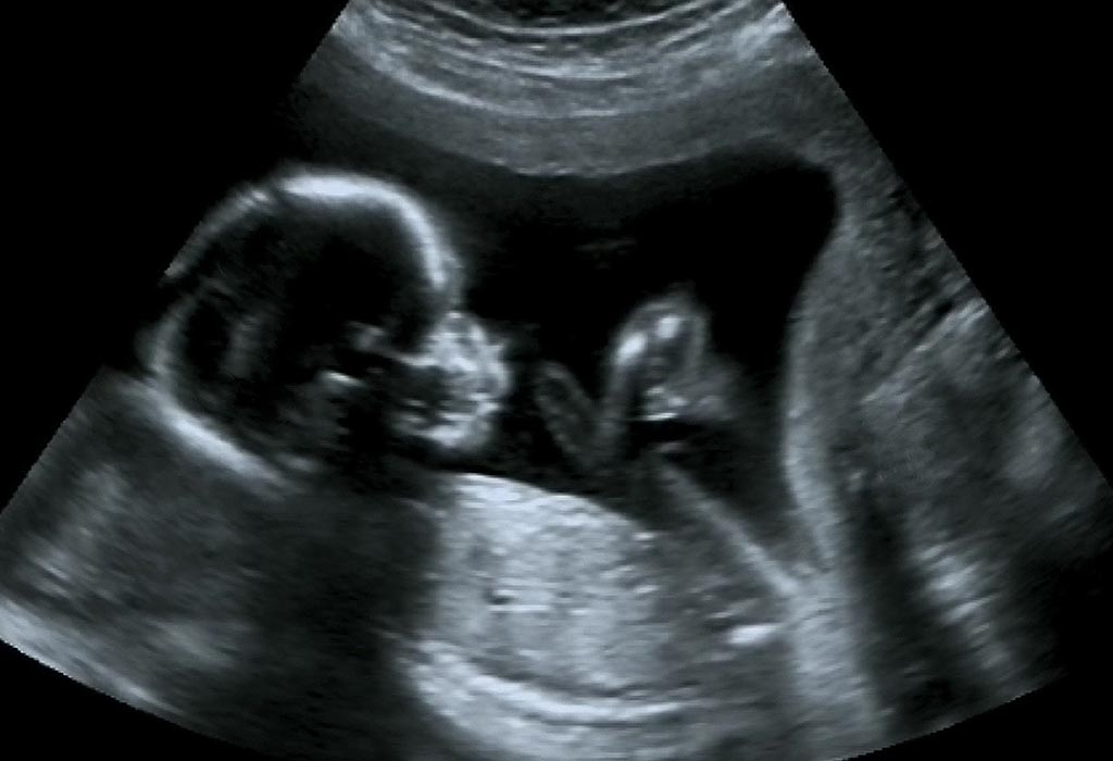
At 18 weeks your baby is growing and moving all around.

5 week ultrasound pictures. Ultrasound pictures by month Credit. 5 weeks 5 days Posted by. Your doctor may recommend more frequent ultrasounds if you have existing health conditions.
10 Week Sonogram Pictures. Our fake ultrasounds are the highest quality on the internet. This can occur but you need to have your physician consider an endocrinology evaluation to be certain.
A 28-year-old female asked. JaesMommy1 1362 views - 1 comment Posted Jun. 5 weeks 5 days Posted by.
A US doctor answered Learn more. Week By Week Ultrasound Pictures. What Can I See on Ultrasound at 5 Weeks.
Ive had one child a son one miscarriage and this is baby number three. We even have 100 authentic and real thermal paper - exactly like you would get at a. If youre 12 weeks along in the pregnancy you may be able to make out your babys head and if youre 20 weeks along you may even see the spine heart feet and eyes.
0 Weeks Pregnant 40 pictures 1 Weeks Pregnant 5 pictures 2 Weeks Pregnant 14 pictures 3 Weeks Pregnant 35 pictures 4 Weeks Pregnant 75 pictures 5 Weeks Pregnant 291 pictures 6 Weeks Pregnant 581 pictures. 5 weeks 3 days Posted by. Babyzzz201 1297 views - 3 comments Posted Jul.
If you are carrying fraternal twins you will be able to see the. Browse Ultrasound Pictures By Week. 6 Week Ultrasound Pictures.
5 weeks 3 days vaginal ultrasound. If you look closely at the image you will see that the labia lips would look similar to a hamburger bun while the clitoris would resemble the hamburger patty. 5 weeks 5 days Posted by.
Dont worry about the numbers and letters at the top of the ultrasound. At 5 weeks your embryo is too small to see yet but the gestational sac may be visible on an ultrasound. 8 Week Ultrasound Pictures.
This is the moniker given to the appearance of the labia and clitoris on an ultrasound. It should be possible for your partner to join you during the ultrasound process and also take a picture of the same although it might cost you. Explore a few of your babys week 5 milestones in this interactive experience.
Baby2018 1143 views - 4 comments Posted Apr. Ursa HoogleGetty Images All ultrasound images for this slideshow were provided by the sonographers of the. This is our 3rd pregnancy.
To read an ultrasound picture look for white spots on the image to see solid tissues like bones and dark spots on the image to see fluid-filled tissues like the amniotic fluid in the uterus. A Verified Doctor answered. If youd like to upgrade from our free version and create an ultrasound with NO watermarks or limitations then check out our Fake Ultrasound shop.
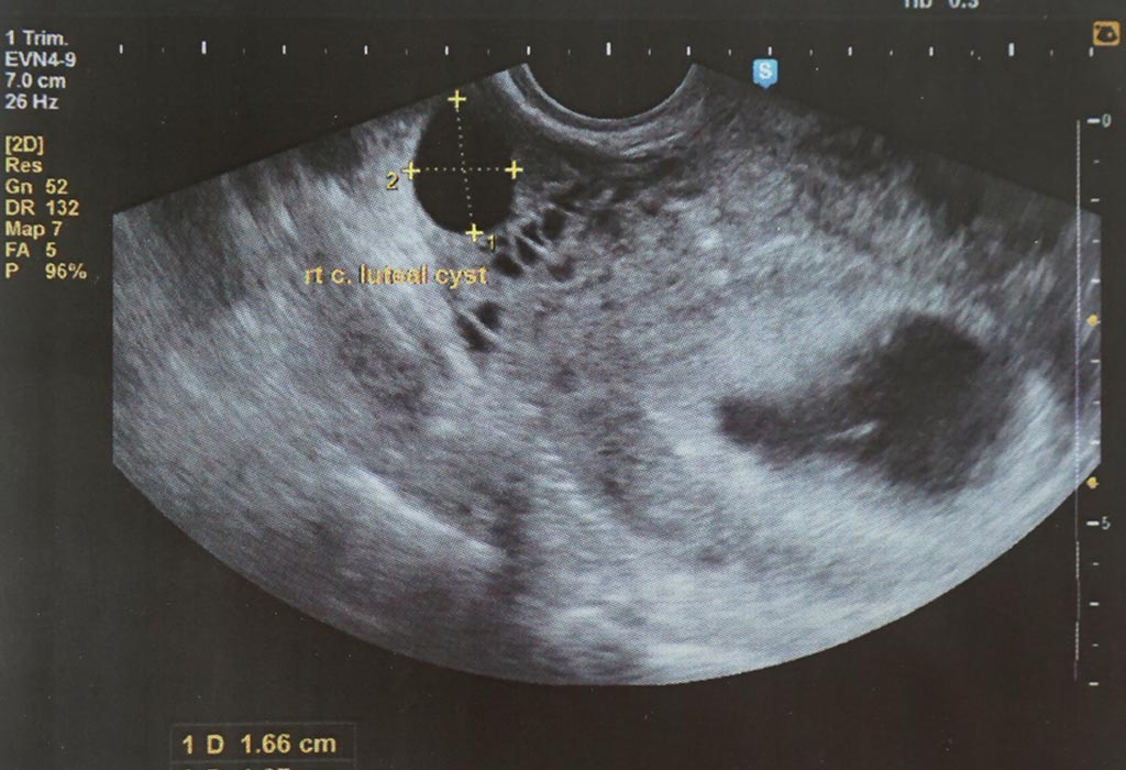
Each sex has a sagittal sign. Five week pregnancy ultrasound with sac and yolk sac Transvaginal ultrasound normal pregnancy at 5 weeks 2 days Gestational sac black area and yolk sac are seen Sac measures 625mm diameter Yolk sac small white circle in left side of the sac Yolk sac is a source of nutrients for the fetus The fetus is too small to be seen this early in pregnancy. If you dont get to peek inside the womb this week youll probably get the chance within the next few weeks.
Our Free Ultrasounds include a Baby Maybe watermark - a small label that shows where the image was generated. At the end of week 5 the heart rate is about 60 90 bpm. In early pregnancies the actual cardiac rate is less important than its presence or absence.
5 Weeks 5 Days Ultrasound. Ultrasound Week By Week. 5 week pregnant ultrasound pictures.
7 Week Ultrasound Pictures. This week you may feel some of those moves and get to see your child demonstrate them on the ultrasound screen. With transvaginal ultrasonography cardiac motion can sometimes be seen in a 2-mm to 3-mm embryo and is invariably detected in normal pregnancy when the length of the embryo reaches 5 mm.
On your 5 weeks pregnant ultrasound you should be able to see your gestational sac and the yolk sac which is always present when you are 5 weeks pregnant. 4 ultrasounds 9 hpts and blood test are all negative but I look five months pregnant and still have pregnancy symptoms why. While most women can expect to see something in a 5-week ultrasound no two pregnancies are the same.
5 Week Sonogram Picture.
I got a redo on my 3D 4d ultrasound since baby boy didnt Cooperate the first time here are some pics of baby 1 at 28 weeks 6 days. However we do recommend a gestational age of 26-34 weeks for the best facial detail.
At 28 weeks babys hearing is so well developed he or she will startle at loud noises.

4d ultrasound 28 weeks pictures. 3D Ultrasound Photos at 14 20 Weeks. Your baby has to hold VERY still so that the high frequency sound waves have time to form around your babys features. First baby pictures Share.
Watch us on Your Carolina. The way 4D3D works is it sends sound waves into the uterus. GoldenView Ultrasound has 3d ultrasound facilities in San Antonio TX New York Cit.
At this stage the baby has put on some weight and filled out to make features more visible yet still enough fluid in front of babys face to obtain great images. See more ideas about 4d ultrasound pictures 4d ultrasound ultrasound pictures. This is baby Kinsley 28 Weeks 4D ultrasound session pictures and video clips.
I love mine and I am TOTALLY happy with doing it. 3D ultrasound photo gallery 14-20 weeks. All 3D ultrasound photos were taken in Greenville SC at Baby.
It is possible you may be scheduled for a more comprehensive and detailed ultrasound. 4d ultrasound Video taken at GoldenView Ultrasound at 28 weeks gestation. Baby Impressions 4D Ultrasound 620 Congaree Road Suite D.
The purpose of this ultrasound is to be sure that your fetus is developing normally. 4-D is similar to 3-D but it shows movement so you can see your baby kicking or opening and closing their eyes. 10 weeks 4 days.
I didnt have to pay for mine though but I would pay for it for sure with my next child if I have to. He was laying the right way to get good facial shots. This ultrasound is often called an anatomy scan.
This looks more like what youre used to seeing in a typical photograph. Baby Impressions 620. In a 3-D ultrasound many 2-D images are taken from various angles and pieced together to form a three-dimensional image.
27 Weeks Pregnant 113 pictures 28 Weeks Pregnant 187 pictures 29 Weeks Pregnant 127 pictures 30 Weeks Pregnant 101 pictures 31 Weeks Pregnant 108 pictures. Even though youd probably love to get a peek inside that 28 weeks pregnant belly its simply not necessary to have more than a couple ultrasounds throughout your pregnancy unless the doctor has a reason to monitor you extra carefully. 14-20 weeks 23-27 weeks 28-32 weeks 33-36 weeks.
Soon baby will starting turning hisher head to listen to sounds outside your body. This is the most beautiful baby in the world and it is the best Christmas gift. Aug 20 2018 - 4D Ultrasound before and after3D Ultrasound PicturesSonogram.
Click photo to enlarge. I had mine done at my last appointment I was 28 weeks and I got two really great pictures before he flipped over. 3D 5D ultrasound images and 4D ultrasound video can be obtained at any stage.
Therefore we suggest getting your babyface ultrasound at 28-34 weeks before baby gets too big and squished into the placenta. If your pregnancy has been uncomplicated dont expect to get a 28-week ultrasound at this appointment. Im only at 31 weeks but I had that 4D3D us and yes baby looked totally deformed.
Recently Posted Ultrasound Pictures. At 28 weeks pregnant a baby typically measures about 10 inches 254 centimeters from the top of their head to the bottom of their buttocks known as the crown-rump length and babys height is over 14 inches 361 centimeters from the top of their head to their heel crown-heel length. It is recommended that you have an ultrasound between 18 to 20 weeks.
28 Week 3d Ultrasound Pictures 28 Week 4d Ultrasound Images 3d 4d Ultrasound Near Me 29 Week 3d Ultrasound Pictures 3d Ultrasound At 28 Weeks. Sounds like you have a very active baby. This special ultrasound is called a level 2 ultrasound and may be recommended if the.
This week the babys weight is about 42 ounces or 2 12 pounds 1189 grams. If your placenta is posterior it will be under the baby and you can wait until 32-38 weeks to have your ultrasound. 11 Week Ultrasound Girl 32 Week 3d Ultrasound Pictures 32 Week 3d Ultrasound.
Large ventricular septal defects. Detection of congenital heart defects throughout pregnancy.

Fetal Heart Ultrasound How To Diagnostic Medical Sonography Ultrasound Obstetric Ultrasound
These ultrasound images suggest a solid non-calcific mass of the pericardium.
Fetal heart defects ultrasound. Sonography of this 36 weeks old fetus revealed a large echogenic mass in close relation to the exterior of the fetal left ventricle within the pericardial cavity. The test helps doctors to see abnormalities in a babys blood flow and. Ventricular Septal Defect or VSD is the most common congenital cardiac defect in children and accounts for 37 percent of cases.
Ultrasound Obstet Gynecol 2006. And with ultrasound most of those defects are caught well before birth giving parents and doctors valuable time to prepare a plan. When such an anomaly is suspected additional fetal malformations should be sought and fetal karyotype should be determined.
Ultrasound in pregnancy enables prenatal diagnosis of CHD which allows for delivery in a facility with appropriate postnatal care. If a heart defect is suspected or a pregnant woman is at risk of having a baby with a heart defect a pediatric or fetal cardiologist will perform a fetal echocardiogram. Large defects are greater than the aortic diameter.
Ostium Secundum Atrial Septal Defect Best visualized on subcostal four-chamber view of the heart. Congenital defects both major and minor occur in around three percent of all births. The examiners ultrasound experience has a significant impact on the detection rate of congenital heart defects at the second-trimester fetal examination.
Congenital heart defects are the most common major structural fetal abnormalities. Wong SF Chan FY Cincotta RB et al. Prenatal ultrasound for detection of fetal anomalies has become a routine part of the pregnancy management in most advanced countries.
Ostium secundum ASDs appear as a larger than expected area of dropout in the central portion of the septum secundum in the vicinity of the foramen ovale or as a deficient foraminal flap septum primum that fails to cover the entire foramen ovale. Situs- check which is the left side of fetus then do a dual image in a tranverse axial plane of the fetus with firstly the thorax showing the heart apex orientated to the left at an angle of approximately 45degrees. Ultrasound Obstet Gynecol 2011.
A small pericardial effusion is also present. Assessment of the four chambers of fetal heart early in pregnancy was feasible and allowed the detection of 45 of CHD. Color flow mapping has played a dominant role in the detection of abnormalities during the first trimester regardless of the International Society of Ultrasound in Obstetrics and Gynecology warning on the use of Doppler during early pregnancy.
First congenital heart diseases CHDs are common congenital anomalies. Should a prenatal ultrasound indicate your baby may have a heart defect or if you have risk factors your obstetrician will most likely order a test called a fetal echocardiogram to examine your babys heart before birth. Possibilities include rhabdomyoma teratoma and hemangioma.
Transvaginal ultrasonography in the early second trimester is a useful tool for the detection of fetal cardiac structural defects provided that both the four-chamber view and the outflow tracts are evaluated. Three- and four-dimensional ultrasound with inversion flow can be used to detect small defects. The goals of the fetal ultrasound diagnosis of VSD are to define whether the segment of the ventricular septal is involved and to.
101002uog8952 The thymicthoracic ratio in fetal heart defects. The accuracy of these tests however is closely related to the stage and type of pregnancy involved. Sound waves ultrasound are used in this test to produce a moving image of the heart.
A fetal echocardiogram echo is a detailed ultrasound exam that takes images of the babys heart. The four chamber view can only detect some of the congenital cardiac anomalies 64 according to one study 2 that can be detected antenatally and these include. M-mode heart rate - should be between 120 and 180 beats per minute.
123 During an assessment where Bi-Plane is used physicians may see what they suspect to be a hole in the fetuss heart. Factors influencing the prenatal detection of structural congenital heart diseases. Congenital heart defects CHD are the most common birth defect with a prevalence of approximately 58 per thousand live births.
Impact of first trimester ultrasound screening for cardiac abnormalities. Of these roughly three out of four will be detected by ultrasound. Fetal cardiac examination is an indispensable part of the prenatal ultrasound because of the following well-recognized reasons.
Detection of fetal VSD is done by cardiac ultrasound performed between 18 and 22 weeks of pregnancy.
Week 17 Week 18 Week 19 Week 20 Week 21 Week 22 Week 23 Week 24 Week 25 Week 26 Week 27 Third Trimester. Hey ladies I was wondering if any of you have had a 3d4d ultrasound done around 16-17 weeks and if baby actually looked like a real baby.

Ivy S 3d Ultrasound 17 Weeks 2 Days Youtube
The big mid-pregnancy ultrasound will most likely take place between 18 weeks and 22 weeks.

17 weeks 3d ultrasound. Experts also discourage the use of any kinds of ultrasounds 2D Doppler 3D and 4D for the purpose of creating a memento. Typically this diagnostic test is performed after 17 weeks. Currently ACOG recommends that expecting women have at least one 2D ultrasound between weeks 18 to 22 of pregnancy noting that some women may also have a first-trimester ultrasound.
All of these images were taken here at SonoSmile which does amazing 3D ultrasound in Ocala Florida 2D 3D and 4D Ultrasound Ocala Florida. The girl ultrasound gallery is designed to show you what a baby girl looks like on ultrasound photos from various weeks of pregnancy. She wasnt very cooperative at first but at 1730 she finally turned around so we could see her entire face.
Due to fetal position and size some of the views at 17 weeks can be suboptimal that means that they are not 100 clear if this is the case a follow up Ultrasound will be recommended at week 20. The purpose of this ultrasound is to be sure that your fetus is developing normally. Our technicians are highly skilled at using the ultrasound technology to our best advantage in determining gender and despite the inherent imperfections in any technology have an unsurpassed accuracy rate.
We often times can determine gender after 15 weeks but we guarantee it after 17 weeks. The average height for a baby at 17 weeks from the top of their head to their heels known as crown-heel length is just under 7 34 inches 196 centimeters. It may be used if amniocentesis results were inconclusive and you and your partner want a more definitive answer about babys health.
18 to 20 Week Ultrasound. The doctor uses an ultrasound to find the place where the cord meets the placentathats the spot where they need to remove the blood. Each week in pregnancy can look slightly different.
Should You Get a Keepsake 3D or 4D Ultrasound. At this stage the baby has put on some weight and filled out to make features more visible yet still enough fluid in front of babys face to obtain great images. Youll notice that what you see varies a lot by the number of weeks of gestation.
At 17 weeks baby measures just over 5 14 inches 135 centimeters when measured from the top of their head to the bottom of their buttocks crown-rump length. 17 week Ultrasound is performed Transabdominally and here is a glimpse of what you can expect to see remember this is the anatomy scan. This ultrasound is often called an anatomy scan.
3D Ultrasound at 17 Weeks or 18. About Press Copyright Contact us Creators Advertise Developers Terms Privacy Policy Safety How YouTube works Test new features Press Copyright Contact us Creators. It is recommended that you have an ultrasound between 18 to 20 weeks.
Second Trimester Ultrasound. This week you may get to see your baby. 3D 5D ultrasound images and 4D ultrasound video can be obtained at any stage.
A 17 weeks pregnant ultrasound may also be recommended to find the cause of vaginal bleeding if any. Hubby got a little too excited tonight and when he was calling for info about a 3d4d ultrasound he actually made an appointment for one day before I am 17 weeks. Im looking into going for a 3D ultrasound before my 20 week ultrasound and Im wondering if its a better experience to go at 17 or 18 weeks.
It is possible you may be scheduled for a more comprehensive and detailed ultrasound. The position of the placenta the condition of the amniotic sac and amniotic fluid the status of the cervix and overall uterine condition can also be checked with the help of a sonogram. However we do recommend a gestational age of 26-34 weeks for the best facial detail.
Ultrasound for the purpose of gender determination is optimal beginning at 17 weeks. Medically reviewed by. Our ultrasound video from 27 weeks.
This page shows typical 3D ultrasound images from 11 to 36 weeks.
Ultrasound week 19 Credit. After this period the babys growth intensifies.

19 Week Pregnant Ultrasound Procedure Abnormalities And More
With the improvements in ultrasound technology the option of a 19-week 3D ultrasound is also available which can provide 3D images of the unborn baby.

19 week ultrasound picture. Anatomy Ultrasound19 weeks and 2 daysa funny storybump picture. Subscribe to receive my posts special offers recipes and. Twin Pregnancy at 19 Weeks.
We are mainly doing it because we want to find out the sex since my regular ultrasound you couldnt tell. 19 weeks 1 day. You should open me.
Length 5 14 to 6 inches crown to rump. Details on 19th week pregnancy symptoms baby development. Their weight reaches 300 grams 10 part of the weight the baby will gain before its birth.
And were you able to see baby good. 19 weeks 1 day. Up until this point in your unborn babys development his head has overshadowed the rest of his body.
19 Weeks Pregnant Ultrasound Pictures. Posted 19 weeks ago. But I was just wondering if the baby is gonna look weird at 19 weeks.
This is the linkwhere u can see the pictureand if u will click on the picture it will enlarge and u can take a proper look please take a look and tell me what do u think. You know you wannaour amazing 4th of july themed firework gender revealhttps. However we do recommend a gestational age of 26-34 weeks for the best facial detail.
I know the facial features wont be great until 26 weeks or so. 19 weeks ultrasound picture boy or a girl. 19 weeks and 2 days baby bump.
The transducer takes a series of images thin slices of the subject and the computer processes these images and presents them as a 3 dimensional image. Their height is approximately 9 inches 228 centimeters from the top of the head to the heel crown-heel length. On 19th week of pregnancy babys height reaches the half of their height during birth 25 cm approximately.
Using computer controls the operator can obtain views that might not be available. During your ultrasound a picture of your baby is produced when high-frequency sound waves bounce off your baby and translate into an image on screen. Repeat 20-week ultrasound because of position of baby.
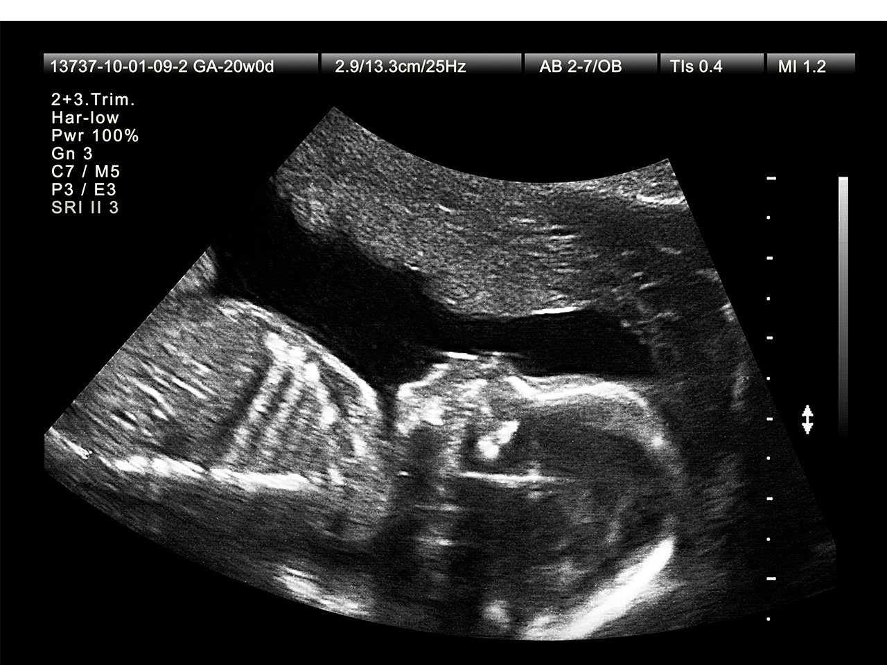
In this image solid matter such as bones are white while softer tissue appears gray. Has anyone gotten one done around 19-20 weeks. Now his arms and.
3D 5D ultrasound images and 4D ultrasound video can be obtained at any stage. Picture 2 is an ultrasound image that shows the foot and leg of a baby girl at 19 weeks 3 days pregnant. 19 week ultrasound.
19 weeks pregnancy update along with photos and an update from our 18 week anatomy ultrasound appointment. In picture 3 as you can see the gender of the baby is revealed in the ultrasound. 235 views - 0 comments.
This is Kims favorite picture from the ultrasound because its a good profile view. 275 views - 0 comments. When I went in for that they could see the spine but still could not get a good picture of the heart so I had to come in a third time a week later.
At 19 weeks your baby measures about 6 14 inches 158 centimeters from the top of their head to the bottom of the buttocks crown-rump length. Picture 1 shows a close up of a baby girl at 19 weeks and 3 days pregnant. To read an ultrasound picture look for white spots on the image to see solid tissues like bones and dark spots on the image to see fluid-filled tissues like the amniotic fluid in the uterus.
I had to go in a second time because they could not get a good picture of the spine or heart. By this week of pregnancy your baby will weigh around 9 12 ounces 272 grams. By this time an unborn baby may also develop some hearing abilities.
At this stage the baby has put on some weight and filled out to make features more visible yet still enough fluid in front of babys face to obtain great images. 3D ultrasound picture of baby at 19 weeks pregnant. Areas that contain fluid such as blood vessels or the stomach as well as the amniotic.
122 views - 2 comments. Your Babys Development at 19 Weeks. The 19-week scan can offer pictures of your unborn baby signifying his growth and development.
If youre 12 weeks along in the pregnancy you may be able to make out your babys head and if youre 20 weeks along you may even see the spine heart feet and eyes.
Halaman
Drbrowns
- January 2022 (32)
- December 2021 (68)
- November 2021 (60)
- October 2021 (70)
- September 2021 (111)
- August 2021 (104)
- July 2021 (93)
- June 2021 (82)
- May 2021 (85)
- April 2021 (102)
- March 2021 (94)
- February 2021 (90)
- January 2021 (86)
- December 2020 (95)
- November 2020 (71)
- October 2020 (101)
- September 2020 (87)
- August 2020 (87)
- July 2020 (99)
- June 2020 (76)
- May 2020 (34)
Label
- 13th
- 17th
- 2015
- 2016
- 4ever
- abortion
- abortions
- about
- abscess
- accurate
- acid
- acorn
- acting
- action
- active
- activities
- actual
- adele
- adoption
- adore
- adults
- advent
- after
- agency
- aggressive
- alisha
- alive
- allergic
- allergies
- allergy
- almond
- alter
- american
- anarchy
- anatomically
- andover
- angelic
- angels
- animal
- animals
- anna
- announce
- announcement
- announcements
- annoyed
- another
- answer
- antibiotics
- applebees
- applegate
- apples
- apraxia
- aquariums
- area
- armpits
- arms
- around
- arrangements
- arrival
- arts
- ashley
- attack
- attitude
- attractions
- austin
- autism
- avent
- aversion
- awake
- babies
- baby
- babys
- babysit
- babysitter
- babysitters
- babysitting
- back
- backpack
- backpacks
- backyard
- bags
- bahama
- baking
- balls
- band
- bangs
- banners
- barbie
- bars
- basal
- baseball
- basketball
- bassinet
- bath
- bathing
- bathroom
- beach
- bean
- beanie
- beans
- bear
- bears
- become
- bedroom
- beds
- bedtime
- beef
- beer
- before
- beginners
- being
- belly
- belt
- benadryl
- bento
- best
- better
- between
- bigger
- bike
- bikes
- binder
- bingo
- biracial
- birth
- birthday
- birthdays
- birthing
- bismol
- bites
- black
- bladder
- blade
- blanket
- blankets
- bleeding
- bloating
- block
- blocked
- blocks
- blood
- bloody
- bloom
- blotches
- blue
- blueberries
- board
- body
- boogers
- book
- books
- boomers
- boost
- booster
- born
- both
- bottle
- bottles
- boundaries
- bowel
- bowling
- bows
- boxes
- boys
- brain
- brands
- bras
- break
- breakfast
- breast
- breastfed
- breastfeeding
- breastmilk
- breathing
- breeze
- brooke
- broth
- brothers
- brown
- browns
- bubble
- budget
- bumbo
- bumpers
- bumps
- bundt
- bunk
- burning
- butter
- butterflied
- butternut
- cabinets
- caffeine
- cake
- cakes
- calculator
- calendar
- california
- call
- calls
- calories
- cameras
- camp
- campfire
- camping
- cana
- cancer
- candy
- canton
- cape
- card
- care
- careers
- carrier
- carrots
- cars
- carving
- casserole
- casting
- cats
- cause
- causes
- cavities
- cell
- cells
- center
- centerpiece
- centerpieces
- cereal
- ceremonies
- cervical
- chair
- chairs
- chamomile
- chances
- change
- changing
- channing
- chart
- cheap
- cheat
- cheating
- checks
- cheerios
- cheese
- chemical
- chest
- chicco
- chicken
- child
- children
- childrens
- chip
- chipmunks
- chives
- chocolate
- choice
- choked
- chokes
- chop
- chores
- christmas
- chuck
- cinnamon
- circus
- classes
- clean
- cleaner
- clear
- click
- climbing
- clinic
- clip
- clippers
- clogged
- closing
- clothes
- clothing
- coach
- coco
- coconut
- coed
- coffee
- coke
- cold
- colic
- college
- color
- coloring
- colors
- colours
- combi
- combinations
- coming
- commercial
- commercials
- company
- conception
- concussion
- cone
- confidence
- congestion
- connected
- cons
- constant
- constipation
- contagious
- contest
- control
- controlled
- convert
- convertible
- cook
- cookies
- cooler
- copper
- cornstarch
- coronado
- correct
- cortisone
- cost
- costa
- costs
- costume
- costumes
- cotton
- cough
- coughing
- country
- couples
- course
- cover
- covered
- craft
- crafts
- cramps
- crawling
- crazy
- cream
- crib
- cribs
- cries
- crockpot
- croup
- crusted
- cuisinart
- cupcakes
- cups
- cure
- curry
- custody
- cute
- cutlets
- cycle
- daddy
- dads
- dairy
- dangers
- dark
- dating
- daughter
- daycare
- days
- dead
- deaf
- deal
- dealing
- deals
- decker
- decor
- decoration
- decorations
- deep
- defects
- definition
- delay
- delivers
- delivery
- dependant
- depo
- depressed
- designs
- desks
- detectors
- development
- deviled
- diabetes
- diagnosis
- diaper
- diapers
- diarrhea
- diastasis
- diego
- dies
- different
- difficult
- dilated
- dirty
- discharge
- disease
- disney
- divorce
- doctor
- does
- doesn
- dogs
- doll
- dolls
- donating
- donation
- donovan
- dosage
- dose
- dots
- double
- down
- drainage
- drawer
- drawers
- drawings
- dream
- dreams
- dress
- dresser
- dresses
- drinking
- drops
- ducks
- duct
- dumpty
- during
- early
- ears
- easter
- easy
- eating
- eczema
- educational
- effaced
- effects
- eggs
- einstien
- elderberry
- elderly
- elegant
- elephant
- elmo
- elsa
- emergen
- emergency
- emiliano
- emotions
- empanadas
- enchiladas
- ended
- episode
- essential
- essentials
- essie
- estrogen
- evenflo
- every
- everyday
- exercises
- exersaucer
- experiments
- exploding
- extended
- extra
- eyes
- face
- faces
- facing
- facts
- fairies
- fairy
- fake
- fall
- families
- family
- farm
- fashioned
- fastest
- father
- fathers
- favors
- feeder
- feeding
- feel
- feeling
- feels
- feet
- felt
- female
- females
- fencing
- fertility
- fetal
- fever
- find
- finger
- first
- fish
- fisher
- fishing
- fitness
- five
- flashes
- floor
- flour
- flower
- focus
- fold
- folding
- food
- foods
- forehead
- formula
- forward
- four
- frappes
- freak
- free
- freeze
- freshly
- friday
- friendly
- friends
- from
- front
- frozen
- fruit
- fudge
- full
- funny
- gain
- game
- games
- gender
- genetic
- gentian
- gentle
- gerber
- getting
- gift
- gifted
- gifts
- gingerbread
- girls
- give
- glazed
- glucose
- gluten
- godzilla
- goes
- going
- good
- graco
- graduation
- graham
- grandma
- grandmother
- grandparents
- gras
- grass
- gravy
- green
- grieving
- grilled
- grippy
- grips
- groin
- groups
- grow
- growth
- gucci
- guide
- guilt
- gummies
- gummy
- guns
- gurgly
- hair
- hallmark
- halloween
- hand
- handle
- handmade
- hands
- hanukkah
- happens
- happiest
- happy
- hard
- hatchable
- hates
- hats
- have
- having
- hayden
- hazel
- head
- healthiest
- healthy
- heart
- heartbeat
- heartbeats
- heating
- heavy
- hebrew
- held
- helmet
- help
- hemorrhoid
- hemorrhoids
- herb
- herbs
- herpes
- hide
- high
- hips
- hitting
- hold
- home
- homemade
- homeopathic
- homes
- honey
- hood
- hopscotch
- hospital
- hostess
- hotels
- house
- hulk
- humpty
- hunt
- hurt
- husband
- hustles
- hydrocortisone
- idea
- ideal
- ideas
- identical
- ikea
- illinois
- images
- implantation
- inappropriate
- inch
- incision
- inclusive
- increase
- incredible
- indian
- indoor
- infant
- infantino
- infants
- infection
- infections
- infertile
- infertility
- inflatable
- ingenuity
- inner
- insect
- inseminate
- installation
- intense
- interactive
- intimate
- into
- invitation
- invite
- invites
- iron
- irregular
- islands
- italian
- italy
- itch
- itchy
- jack
- jacket
- james
- jealous
- jeans
- jessie
- jewish
- jobs
- joes
- jude
- juice
- july
- jungle
- just
- justin
- jwoww
- karts
- kate
- keep
- kelty
- kids
- kindergarten
- kindergartners
- kitchen
- kits
- know
- korean
- kristoff
- labels
- labor
- ladder
- ladies
- lake
- lamb
- language
- large
- laser
- last
- late
- laugh
- leader
- learn
- leave
- leaved
- leaving
- left
- lego
- legs
- lenox
- lesson
- letters
- leukemia
- levels
- lice
- life
- lift
- lifting
- ligation
- light
- lights
- lightweight
- like
- line
- lips
- liquid
- list
- little
- live
- living
- lock
- locks
- long
- looks
- lose
- loss
- lotrimin
- loud
- louis
- love
- lower
- lubricants
- lullaby
- lump
- lumps
- lyme
- macaroni
- machine
- made
- make
- makeover
- makes
- makeup
- male
- mama
- mango
- manual
- many
- mardi
- marinade
- marisol
- mark
- marks
- marmalade
- mashed
- mask
- matching
- maternity
- maths
- mcstuffins
- meal
- meals
- mean
- meaning
- meat
- medela
- medicine
- meditation
- medula
- melatonin
- memorial
- menstrual
- menu
- mermaid
- mesh
- message
- metairie
- metal
- michaels
- mickey
- midwife
- mild
- milestones
- milk
- mini
- miniature
- minnie
- miracle
- mirena
- mirror
- mirrors
- miscarriage
- moana
- moccasins
- model
- mommy
- moms
- monistat
- monitor
- monitoring
- monitors
- monkey
- montego
- month
- months
- morning
- moses
- mosquito
- most
- mother
- mothers
- motion
- motor
- mountain
- mouse
- mouth
- move
- movement
- movie
- movies
- moving
- much
- mucus
- muffin
- multivitamin
- munchkin
- museum
- museums
- music
- musical
- mustard
- name
- names
- naming
- nanny
- napkins
- native
- natural
- nausea
- nautical
- navy
- neck
- necklace
- necklaces
- needs
- negative
- neti
- neutral
- newborn
- newborns
- newspaper
- next
- niÃo
- niece
- night
- nightgown
- nightgowns
- nike
- ninja
- nits
- noodles
- nose
- note
- notes
- nuggets
- numb
- number
- nursery
- nurses
- nursing
- nyquil
- occupational
- offenders
- ohio
- oils
- older
- oldest
- olds
- omeprazole
- open
- opening
- optical
- oral
- orange
- organic
- organics
- organizing
- origin
- ornament
- ornaments
- other
- ottoman
- outdoor
- outfit
- over
- overnight
- ovulation
- pacifiers
- pack
- packing
- pads
- pages
- pain
- painful
- paint
- painting
- pajamas
- panic
- pants
- paper
- parent
- parenting
- parents
- park
- part
- parties
- party
- pass
- pasta
- peanut
- pear
- pedialyte
- pensacola
- people
- pepcid
- pepper
- peppers
- period
- periods
- phil
- phone
- photo
- photos
- physical
- pick
- picked
- picture
- pictures
- piece
- pill
- pillow
- pills
- pillsbury
- pineapple
- pink
- pirates
- pizza
- place
- places
- plan
- planning
- plans
- plaque
- plastic
- plates
- play
- player
- playhouses
- playing
- playpens
- plus
- pneumonia
- poem
- poison
- polish
- polycystic
- pony
- pooh
- pool
- popcorn
- popular
- pork
- portable
- poster
- postpartum
- potato
- potatoes
- potty
- pounds
- powder
- prediction
- pregnancy
- pregnant
- premature
- prenatal
- preparing
- preschool
- preschoolers
- preschools
- prescription
- present
- presents
- pressure
- prevent
- price
- printable
- printables
- printing
- prodromal
- products
- projects
- pros
- protection
- protein
- providers
- psychological
- psychology
- ptosis
- pudding
- puerto
- pull
- pulled
- pump
- pumping
- pumpkin
- punta
- push
- qualifications
- questions
- quinoa
- quotes
- rachael
- rachel
- racism
- rail
- rainforest
- raising
- range
- rash
- rashes
- raspberry
- rate
- rates
- reaction
- reading
- real
- rear
- reasons
- recall
- recalled
- recalls
- recipe
- recipes
- reduce
- reflux
- reglan
- relationship
- relationships
- relaxation
- relief
- religion
- remedies
- remedy
- remote
- removal
- removing
- reno
- repellent
- replacement
- resorts
- respect
- restaurant
- restaurants
- reusable
- reveal
- reviews
- ribs
- rice
- ride
- rider
- riding
- right
- ring
- risk
- risks
- roast
- roasted
- robe
- robes
- robitussin
- rock
- rocket
- rocking
- roll
- rolling
- rolls
- romance
- ronald
- room
- rooms
- root
- rosemary
- round
- rubber
- rubiks
- runny
- sack
- safe
- safety
- saftey
- salad
- sale
- saline
- salmon
- salt
- sandwiches
- sanitizer
- santa
- sauce
- sauerkraut
- sausage
- scab
- scanner
- scary
- scavenger
- schedule
- schedules
- schools
- science
- scissors
- scooby
- screen
- search
- seat
- seater
- seats
- section
- seed
- selection
- self
- sensation
- sensitive
- sensor
- setting
- sexual
- shampoo
- shaped
- shapes
- sharing
- sheet
- shirt
- shirts
- shoes
- shoot
- short
- shortcake
- shot
- shots
- should
- show
- shower
- showers
- showing
- siblings
- sick
- sickness
- side
- sign
- signs
- sinus
- sippy
- sister
- site
- size
- skid
- skin
- skins
- skirt
- sleds
- sleep
- sleeper
- sleeping
- slide
- sling
- small
- smart
- smell
- smells
- smoking
- snapchat
- snow
- snowsuit
- socks
- soda
- soft
- solids
- some
- someone
- song
- songs
- soon
- sore
- sound
- sources
- south
- southern
- space
- spacesaver
- spade
- spaetzle
- special
- speech
- spice
- spinach
- spit
- spitting
- spoil
- sports
- spotting
- spout
- spray
- springbreak
- springform
- squash
- stages
- stand
- standing
- star
- starbucks
- start
- starting
- statistics
- stay
- steak
- step
- sterling
- stick
- stickers
- stiff
- stocking
- stockings
- stokke
- stomach
- stool
- stools
- stop
- stories
- stork
- story
- strap
- straps
- straw
- strawberry
- strech
- strep
- stretch
- stroller
- strollers
- strong
- student
- stuff
- stuffed
- stuffer
- style
- successful
- sugar
- suit
- suits
- summer
- sunburn
- sunscreen
- super
- superhero
- supplementing
- supplies
- supply
- support
- surrogate
- suvs
- swaddle
- sweater
- sweaters
- sweating
- sweats
- sweet
- swim
- swimming
- swimsuits
- swing
- swings
- switch
- symbols
- sympathy
- symphony
- symptom
- symptoms
- syndrome
- syrup
- system
- systems
- table
- taffy
- tail
- tails
- take
- tales
- talking
- talks
- tampa
- taping
- tattoos
- teach
- teacher
- teachers
- teaching
- tear
- tech
- techniques
- teddy
- teds
- teen
- teenage
- teenager
- teenagers
- teens
- teeth
- teether
- teething
- tell
- telling
- tells
- temper
- temperature
- templates
- tent
- tents
- test
- testing
- thank
- thanksgiving
- that
- their
- theme
- themed
- themes
- themselves
- therapists
- therapy
- there
- thighs
- thing
- things
- third
- this
- throat
- thrones
- throwing
- thrush
- tick
- ticks
- tight
- tike
- tikes
- time
- timer
- tiny
- tips
- toddler
- toddlers
- toilet
- tomato
- tonight
- tooth
- toothbrush
- tops
- toxic
- toys
- trade
- trader
- trainer
- training
- trampoline
- transgender
- transition
- travel
- travelling
- treat
- treating
- treatment
- treatments
- trek
- trend
- trick
- trimester
- trimmers
- troubled
- truck
- true
- trunks
- trying
- tubal
- tubes
- tums
- tuna
- turkey
- turtle
- tutu
- tweens
- twin
- twins
- twitching
- tylenol
- types
- ugly
- ultrasound
- umbilical
- unborn
- uncircumcised
- under
- unicorn
- unique
- unscramble
- upgrade
- uppababy
- upper
- upset
- urine
- used
- uterus
- vacation
- vacations
- vaccinate
- vaccine
- valentine
- valentines
- vanilla
- vanishing
- vegans
- vendors
- video
- videos
- violet
- virgin
- visit
- vitamin
- vomiting
- vote
- waist
- walker
- walkers
- walking
- wall
- wallpaper
- walmart
- want
- wants
- warm
- warmer
- warrior
- wars
- warts
- wash
- watch
- water
- wean
- wear
- wearable
- wedding
- weddings
- week
- weeks
- weight
- whale
- what
- when
- where
- while
- white
- whole
- whooping
- wife
- will
- willed
- wings
- winter
- witch
- with
- without
- wizard
- wolf
- woman
- women
- womens
- wont
- wooden
- woods
- wording
- words
- work
- working
- workouts
- worksheets
- world
- would
- wrap
- wraps
- wyatt
- yard
- yards
- year
- years
- yeast
- yellow
- yogurt
- york
- your
- yourself
- zantac
- zoom
- zyrtec

